Video

Sunday, 30 April 2017

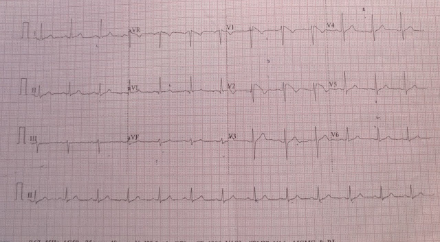
Atrial flutter with complete heart block

A 51-year-male came to JIPMER Pondicherry, India with chief complaints of giddiness since last one week. There were no history of syncope, palpitation, chest pain. There was no past history of diabetes mellitus, hypertension. ECG of the patient is shown below

ECG 1(Click on the image to enlarge it)


ECG is suggestive of heart rate of 38 beats per minute, atrial rate around 300 beats per minute, saw tooth P wave seen in lead V1, there is no definite baseline seen between P wave, P wave with saw tooth were negative in  lead 2,3,avf suggestive of atrial flutter. The heart rate is nearly regular, no variability seen so the possibility in this patient is either Atrial flutter with block  (6:1) (Counterclockwise  typical flutter) or  an underlying complete heart block. 

Another ECG of the same patient (ECG 2)


Patient underwent EPS study and found to have complete heart block with atrial flutter arising from right atrium. Patient underwent successful radiofrequency ablation. In view of his Complete heart block he latter underwent successful permanent pacemaker implantation.

Thank you.

Typical atrial flutter (AFL) is a macroreentrant atrial tachycardia (AT) that uses the cavotricuspid isthmus as an essential part of its circuit. The circuit boundaries are the tricuspid annulus, crista terminalis, IVC, eustachian ridge, CS os, and probably the fossa ovalis. These barriers (lines of
conduction block) can be functional or anatomical, and are necessary to provide adequate path length for the flutter reentry circuit. The tricuspid annulus forms the anterior border of the flutter circuit,
whereas the posterior border occurs at a variable distance from this anterior border; it is narrowest in the region of the eustachian ridge and widest in the anterior part of the RA. Typical AFL is of two types, counterclockwise and clockwise.2 In counterclockwise AFL, activation proceeds caudocephalic up the septal side of the tricuspid annulus toward the crista terminalis and moves cephalocaudal along the lateral wall of the RA to reach the lateral tricuspid annulus, after which it propagates through the isthmus defined by the IVC, CS, and tricuspid annulus (counterclockwise as viewed in the left anterior oblique [LAO] view from the ventricular side of the tricuspid annulus). The circuit is entirely in the RA. In clockwise (reverse typical) AFL, activation propagates
in the opposite direction

ELECTROCARDIOGRAPHIC FEATURES
Typical Atrial Flutter P Waves. Flutter waves appear as atrial complexes of constant morphology, polarity, and CL. Typically, flutter waves are most prominent in the inferior leads (II, III, aVF)
and V1. In the inferior leads, they appear as a picket fence varieties coexist with tall positive, small positive, or biphasic (sawtooth) because the leads are primarily negative. This consists of a downsloping segment, followed by a sharper negative deflection, and then a sharp positive deflection,
with a positive overshoot leading to the next downsloping plateau. The relative size of each component can vary markedly. Counterclockwise AFL  can be characterized by pure negative deflections in the inferior leads, negative and then positive deflections that are equal in size, or a small negative and then a larger positive deflection. Those three P waves in V1, respectively. The degree of positivity in the inferior leads appears to be related to the coexistence of heart disease and LA enlargement. Counterclockwise AFL will always have a negative deflection preceding the positive deflection in the inferior leads. Leads I and aVL characteristically show low-voltage deflections. Clockwise AFL generally has broad positive deflections in the inferior leads and wide negative deflections in V1.

Reference
Issa Z, Miller JM, Zipes DP. Clinical Arrhythmology and Electrophysiology: A Companion to Braunwald's Heart Disease: Expert Consult: Online and Print. Elsevier Health Sciences; 2012 Apr 24.

Saturday, 15 April 2017

Ventricular tachycardia


A 53-years-old female came to JIPMER, Pondicherry, India with chief complaints of dyspnoea on exertion class III since last five days along with palpitation, perspiration. There was no complaints of chest pain or angina.Patient was a known case of diabetes mellitus, hypertension since last five years and she was on medical treatment for the same. There was no history of coroanry artery disease in the past. ECG of the patient done in the emergency department is shown below.

ECG 1(Click on the image to enlarge it)


Thank you

Tuesday, 11 April 2017

Brugada ECG

A 50year old male came to JIPMER Pondicherry, India with chief complaints of atypical chest pain on and off since last one week. There was no history of giddiness, syncope, palpitation, family history of sudden cardiac death. There was no past history of diabetes mellitus, hypertension, coronary artery diseases. ECG of the patient is shown below.ECHO heart of the patient was normal.

ECG 1


Normal sinus rhythm at 75 beats/min, normal axis,  there is rSr pattern seen in lead V1, no other ST-T wave changes seen, PR interval 120 msec, QT interval 360 msec


Patient ECG repeated after 10 minute

ECG 2

ECG is showing ST-segment elevation with T wave inversion in lead V1, V2. ST-segment elevation in lead V1 is 2 mm, in lead V2 is 4 mm

ECG 3

ECG after 20 minute shows ST segment elevation of 3 mm in lead V1 and 5 mm in lead V2


ECG 4


ECG after 30 minute shows ST segment elevation of 3 mm in lead V1 and 5 mm in lead V2


ECG 5


ECG is showing ST elevation in lead V1,V2

ECG 6


ECG is showing ST elevation in lead V1,V2

ECG of the patient was suggestive of incomplete RBBB with Type I Burgada pattern of ECG.

Cardiac marker of the patient were negative. TMT of the patient , there was no evidence of inducible ischemia.

So on the basis of ECG patient diagnosis is Brugada syndrome (asymptomatic). Because patient was asymptomatic so patient was advised to follow up in cardiology OPD of JIPMER hospital.


Brugada syndrome


Emedicine

Brugada syndrome is a disorder characterized by sudden death associated with one of several ECG patterns characterized by incomplete right bundle-branch block and ST-segment elevations in the anterior precordial leads.


Brugada syndrome has three different ECG patterns (Wikipedia)

  • Type 1 has a coved type ST elevation with at least 2 mm (0.2 mV) J-point elevation and a gradually descending ST segment followed by a negative T-wave.
  • Type 2 has a saddle-back pattern with a least 2 mm J-point elevation and at least 1 mm ST elevation with a positive or biphasic T-wave. Type 2 pattern can occasionally be seen in healthy subjects.
  • Type 3 has either a coved (type 1 like) or a saddle-back (type 2 like) pattern, with less than 2 mm J-point elevation and less than 1 mm ST elevation. Type 3 pattern is not rare in healthy subjects.


Signs and symptoms

Signs and symptoms in patients with Brugada syndrome may include the following:
  • Syncope and cardiac arrest: Most common clinical manifestations; in many cases, cardiac arrest occurs during sleep or rest
  • Nightmares or thrashing at night
  • Asymptomatic, but routine ECG shows ST-segment elevation in leads V1-V3
  • Associated atrial fibrillation (20%) [1]
  • Fever: Often reported to trigger or exacerbate clinical manifestations
The lack of a prodrome has been reported to be more common in patients with ventricular fibrillation documented as the cause of syncope in patients with Brugada syndrome.

Diagnosis

Most patients with Brugada syndrome have a normal physical examination. However, such an examination is necessary to exclude other potential cardiac causes of syncope or cardiac arrest in an otherwise healthy patient (eg, heart murmurs from hypertrophic cardiomyopathy or from a valvular or septal defect).
Testing
In patients with suspected Brugada syndrome, consider the following studies:
  • 12-lead ECG in all patients with syncope
  • Drug challenge with a sodium channel blocker in patients with syncope without an obvious cause
  • Electrophysiologic study to determine the inducibility of arrhythmias for risk 

    Management

    To date, the only treatment that has proven effective in treating ventricular tachycardia and fibrillation and preventing sudden death in patients with Brugada syndrome is implantation of an automatic implantable cardiac defibrillator (ICD)

Thank you.


Monday, 10 April 2017

Wellen"s syndrome

A60 years old male, came to JIPMER,Pondicherry, India with chief complaints of chest pain on and off since last month which increases on exertion. Patient came to JIPMER hospital one month back, ECG done at that time is shown below. 

ECG 1(Click  on the image to enlarge it)


ECG is showing normal sinus rhythm at 64 beats per minute, normal axis, biphasic T wave seen in lead V2-V4, T wave inversion seen in lead V5,V6. 
Patient cardiac marker were negative. So diagnosis of Acute coronary syndrome with unstable angina was made. Patient was planned for coronary angiography planned but could not be done due to logistic reason. Patient went home and again come back to JIPMER hospital emergency with acute onset retrosternal chest pain since one week, increased since 3 hours of duration. ECG of the patient done is shown below
ECG 2(Click on the image to enlarge it)


ECG is showing normal sinus rhythm at 88 beats per minute, normal axis, normal PR interval,  Biphasic T wave with Deep T wave inversion seen in lead V2-V4, ST segment depression with T wave inversion seen in lead V5,V6, No evidence of Q wave anywhere or any poor R wave progression so the ECG is suggestive of classical Wellen"s syndrome.

ECG 3( Done after two hours of hospital  admission)


ECG after 2 hours is showing increase in T wave inversion in lead V2-V6, loss of biphasic T wave in lead V2-V4, no Q wave or no poor R wave progression

ECG done again after 1 minute of the above ECG


ECG is showing T wave inversion in lead V2-V6

ECG done two days after admission to the hospital shown below


ECG is still showing T wave inversion in lead V2-V6


Patient cardiac markerwas done and troponin I was found to be elevated. Coronary angiography of the patient showed proximal left anterior descending artery occlusion ( 90% ). Patient was planned for percutaneous coronary intervention and he is awaiting procedure.


Little about Wellen's syndrome

Wellen's syndrome was first described by Gerson and colleagues in 1980 as an inverted U-wave, and again in 1982 by De Zwaan, Wellens and colleagues as Wellen's syndrome.
 It consists of a characteristic EKG finding suggesting severe stenosis of the proximal LAD artery, which will develop into an acute anterior wall MI within a few days to weeks in 75% of untreated patients.
Wellen's syndrome is diagnosed based on the classic T-wave findings seen on an EKG taken when the patient is pain-free. These T-wave changes represent reperfusion of the myocardium.
There are two variations of Wellen's syndrome T-wave. Type A is the more common abnormality, occurring in 75% of cases, and is characterized by deeply inverted T-waves in V2 and V3. Type B occurs in 25% of cases and is characterized by biphasic T-waves in V2 and V3.
The diagnostic leads for T-waves of Wellens' syndrome are V2 and V3, corresponding with a lesion between the first and second septal branches of the LAD. However, if the lesion is more proximal in the LAD, the T-wave changes will be more widely spread along the precordial leads.
An EKG obtained during episodes of pain will demonstrate upright T-waves with possible ST segment elevation or depression, but an isoelectric ST segment may also be seen.  Cardiac enzymes will be normal or mildly elevated. 
 These changes are easily missed, and therefore it is critical for the Emergency Physicians to be aware of them. It is unlikely that any Emergency Physician would miss the deeply inverted T-waves that occur in this syndrome when the patient is pain-free, as is shown in our case, but the significance of these findings must also be recognized. Emergency angiography is justified with either of these EKG presentations, with the hope of avoiding an extensive anterior wall MI through early intervention.[
Wellens syndrome is also referred to as LAD coronary T-wave syndrome. Syndrome criteria include the following:
    • Characteristic T-wave changes
    • History of anginal chest pain
    • Normal or minimally elevated cardiac enzyme levels
    • ECG without Q waves, without significant ST-segment elevation, and with normal precordial R-wave progression
    Recognition of this ECG abnormality is of paramount importance because this syndrome represents a preinfarction stage of coronary artery disease (CAD) that often progresses to a devastating anterior wall MI
  • Thank you
Reference
De Zwaan C, Bar FW, Wellens HJ. Characteristic electrocardiographic pattern indicating a critical stenosis high in left anterior descending coronary artery in patients admitted because of impending myocardial infarction. Am Heart J. 1982;103:730–6. [PubMed]
2. Movahed MR. Wellen's Syndrome or Inverted U-waves? Clin Cardiol. 2008;31:133–4. [PubMed]
3. Smith S, Whitwam S. Acute Coronary Syndromes. Emerg Med Clin N Am. 2006;24:53–89. [PubMed]
4. Nisbet B, Zlupko G. Repeat Wellen's syndrome: Case report of critical proximal left anterior descending artery restenosis. J Emerg Med. 2008. [PubMed]
5. Sobnosky S, Kohli R, Bleibel S. Wellen's Syndrome. Int J Cardiol. 2006;3:1.
6. Hovland A, Bjomstad H, Staub U, Vik-Mo H. Reversible ischemia in Wellen's syndrome. J Nucl Cardiol. 2006;13:13–5. [PubMed]
7. Tandy TK, Bottomy DP, Lewis JG. Wellen's syndrome. Ann Emerg Med. 1999;33:347–51. [PubMed]
8. Wellens HJJ, Conover MB. The ECG in emergency decision making. WB Saunders Company. 1992. p. 32.
9. Elmenyar A. Wellens Syndrome. Heart Views. 2000-2001;1:408–10.

Sunday, 9 April 2017

Posterior wall myocardial infarction with inferior wall infaction

A 47 years old male came to JIPMER, Pondicherry, hospital with chief complaint of acute onset retrosternal chest pain since 1 day along which was heaving in nature . There was no history of  palpitation perspiration, giddness. Patient was a known case of diabetes mellitus since 15 years and was on oral hypoglycemic agent. Patient ECG is shown below

ECG (Click on the image to enlarge it)


ECG is showing normal sinus rhythm at 84 beats per minute, there is ST segment depression seen in lead I,avL, V2. There is high take off of ST segment seen in lead 2,3,aVF. Prominent R wave seen in lead V1,V2,V3.

ECG of the patient repeated after 10 minute


ECG is showing normal sinus rhythm at 120 beats per minute, right axis deviation, no ST-T wave changes seen.


In view of continuou chest pain ECG with posterior lead was taken

ECG with posterior lead


ECG is showing ST segment elevation in lead V7,V8,V8 which is only 2 small boxes or in other word less than 0.5 mm, there were high take off of ST segment seen in lead 2,3,avF.

Again ECG of the patient was repeated after 6 hours which is shown below


ECG is showing QS complexes in lead 2,3,avF. So the diagnosis of the patient is inferior wall myocardial infarction with posterior wall myocardial infarction. Patient underwent angiography which was suggestive of LAD 70% stenosis, LCX 90% stenosis, RCA 70% stenosis.


Thank you.


PSVT

A 48 year old female came to JIPMER Hospital, Pondicherry, India with chief complaints of palpitation on and off since last one year. There was no history of dyspnoea, chest pain, giddiness, syncope, or any other systemic illness. ECG of the patient done during the episode of palpitation is shown below.
                                           ECG 1(Click on the image to enlarge it)

 ECG is showing narrow complex regular tachycardia at rate around 180 beats/min, normal axis, no ST-T wave seen, no visible P wave seen, normal axis, so the differential is AVNRT


                                    Another ECG of the same patient during tachycardia


Diagnosis is narrow complex regular tachycardia differential being AVNRT.

Approach in a patient with narrow complex regular tachycardia


Thank you.