Video

Thursday 26 January 2017

Ventricular Tachycardia Originating From the Posterior Papillary Muscle in the Left Ventricle

A 53 year old  female resident of Thiruvannamalai, Tamilnadu came to JIPMER hospital emergency with  chief complaints of 10-12 episodes of generalised tonic-clonic convulsion along with transient loss of consciousness since one day. Loss of consciousness remain for 5-10 seconds and patient regain consciousness on its own. These episode were associated with frothing from the mouth and urinary incontinence. Patient was a known case of diabetes mellitus and hypertension since last 5 years and was on regular treatment. There was no history of coronary artery diseases, tuberculosis or any other major illness. During evaluation in the emergency department patient pulse rate was 80 per minute, irregular, Blood pressure was 130/80 mmhg, patient  was conscious , cooperative and oriented to time, place, person, there was no focal neurological deficit, Cardiovascular system S1S2 were present, respiratory system bilateral normal vesicular sound were present. ECG of the patient done in the emergency department is shown below.

                                                   ECG 1(Click on the image to enlarge it )


Description of the ECG-Broad complex tachycardia with QRS of RBBB morphology, QRS duration 140 msec,  left axis deviation, P wave are present merged into the QRS complex suggestive of Atrioventricular dissociation, intermittent capture beats are present with normal PR interval, narrow QRS complex, normal axis of sinus beat, so the ecg is suggestive of ventricular tachycardia with RBBB morphology suggestive of origin from the left ventricle.


ECG during sinus rhythm 2


ECG Sinus rhythm at 115 beats per minute, normal axis, PR interval 160 msec, narrow QRS complex, T wave inversion with ST segment flattening present in lead II,III,aVF, V4-V6, Intermittent VPC present with RBBB morphology with left axis deviation with rS in lead V6 present.

So on the basis of above ECG finding patient cardiac marker were sent (Troponin I ) ,which comes within normal limit. Patient echocardiogram done which was suggestive of global hypokinesia of left ventricle with moderate to severe mitral regurgitation, moderate tricuspid regurgitation was present with right ventricular systolic pressure of 40 mmhg. Patient underwent holter monitoring which was suggestive of  60,000 thousands PVC, runs of ventricular tachycardia with RBBB morphology with left axis deviation with rS morphology in lead V6. Patient underwent Coronary angiography which was suggestive of normal coronary. 
So the diagnosis of Ventricular tachycardia ??Left posterior fasicular tachycardia  ??? Left posterior papillary muscle tachycardia was kept. Patient was planned for electrophysiological study. Patient underwent EPS study which was suggestive of earliest ventricular activation during the VPC close to left ventricle apex close to inferior septum, There was no presystolic purkinje potentials and echocardiogram was suggestive of proximity of this location to the posterior papillary muscle. Pace Mapping  from this site showed a 11/12 pace match. Patient underwent successful CARTO guided ablation and later discharged in stable condition.

So the final diagnosis was Idiopathic Left posterior papillary muscle Ventricular tachycardia with tachycardia induced cardiomyopathy.

Little about Idiopathic Left posterior papillary muscle Ventricular tachycardia 

VT localized to the base of the Posterior papillary muscle  (PPM) in the LV characterized by: (1) a normal baseline ECG and intracardiac conduction intervals with normal LV systolic function; (2) right bundle-branch block and superior-axis QRS morphology; (3) lack of inducibility with programmed ventricular and atrial stimulation; (4) absence of criteria for transient entrainment; (5) inducibility of VT or PVCs with intravenous isoproterenol or epinephrine; (6) earliest ventricular activation at the base of the PPM in the LV; and (7) absence of high-frequency potentials at the site of origin, which suggests that the Purkinje system is not directly involved. Successful catheter ablation at this site uniformly required the use of cooled RF ablation, which suggests that the site of origin was within the papillary muscle itself, somewhat deep within the myocardium.

Reference
Doppalapudi H, Yamada T, McElderry HT, Plumb VJ, Epstein AE, Kay GN. Ventricular Tachycardia Originating From the Posterior Papillary Muscle in the Left VentricleCLINICAL PERSPECTIVE. Circulation: Arrhythmia and Electrophysiology. 2008 Apr 1;1(1):23-9.


Thank  you.


Wednesday 25 January 2017

Middle age female with sinus node dysfunction

A 43 years old female resident of Villupuram, Tamilnadu came to JIPMER Hospital Cardiology OPD with  chief complaints of atypical chest pain since last 6 month, which was left side in location, severe in intensity, occur on and off , was not associated with any palpitation, vomiting, radiation or refernce of pain or any reliving or aggravating factor. Patient was also giving history of dyspnoea on exertion on and off since last 6 month but there was no history of orthopnoea or paroxysmal nocturnal dyspnoea.Patient was a known case of diabetes mellitus and hypertension since the past 2 years and she was on oral medication. There was no  history of beta blocker intake for the hypertensionn. During evaluation in the OPD patient pulse was 60 beats per minute, blood pressure was 100/60 mmhg. Cardiovascular system S1S2 were present, Respiratory system Bilateral normal vesicular sound were present. ECG of the patient done which  is shown below.ECHO heart suggestive of moderate tricuspid regurgitation with right ventricular systolic pressure 42 mmhg. 

ECG 1(Click on the image to  enlarge it)


Description of the ECG-Heart rate 60 beats per minute, Sinus rhythm present with rate around 75 beats per minute, narrow QRS complex, QT interval 460 msec, Corrected QTc interval 510 msec, there were no ST-T wave changes seen, intermittent long sinus pause are present with junctional escape beats with narrow QRS morphology with retrograde P wave merged into the end of QRS complex so on the  basis of this ECG provisional diagnosis was Symptomatic sinus bradycardia.

 Patient underwent holter monitoring for further conform the diagnosis. Holter strips are given below.

Holter strip 1


                    Holter is showing long sinus arrest, sinus pause in above strip was 3.6 second

Holter strip 2


Holter showing long sinus pause with maximum interval being 2.878

Holter strip 3


Holter showing long sinus pause with maximum interval of sinus pause being 1.8 second.

So in the view of sinus pause and history of breathless diagnosis of symptomatic sinus bradycardia was kept. Patient was implanted permanent pacemaker and she was later discharged in stable condition. 

Thank you.


Monday 23 January 2017

5 year old child with congenital Complete heart block

A 5 years old female child resident of Madurai, Tamilnadu, came to JIPMER hospital emergency  with chief complaints of giddiness followed by loss of consciousness four days back, which was sudden in onset associated with one episode of vomiting but there is no history of palpitation, fever, joint pain, anorexia or weight loss. For all these complaints patient went of local goverment hospital, where ECG done suggestive of bradycardia, so patient was referred to JIPMER hosptial for further management. During evaluation in the emergency patient pulse was 50 beats per minute, BP-100/60 mmhg, ECHO done suggestive of dilated left ventricle with LVEF around 50%. ECG of the patient done is shown below.

                        ECG 1(Click on the image to enlarge it )


Descriptionn of the ECG-Complete AV dissociation seen, atrial rate 100 beats per minute, ventricular rate 50 beats per minute, QRS duration prolonged around 160 msec, LBBB morphology,QT interval 520 msec, QTc interval 475 msec, Deep T wave inversion seen inn lead I, II, avF, V2-V6, so the diagnosis of the patient is congenital complete heart block.


                                                                 ECG 2


Another ECG of the patient again showing Complete AV dissociation suggestive of complete heart block. In view of absence of any secondary causes and age of the patient (5 years) the diagnosis of congenital heart block was kept.

Patient was put on permanent pacemaker and was discharged from the hospital in  stable condition.

Thank you.


Saturday 21 January 2017

Varying degree of AV block in old lady

A 64 years old female resident of pondicherry (South India) came to JIPMER emergency with chief complaints of giddiness since last one day along with atypical chest pain. There was no histroy of syncope, palpitation, dyspnoea, vomiting. Patient was a known  case hypertension since last one six years and was on tablet metoprolol 50  mg and tablet telmisartan 25 mg once a day. Patient was also giving history of diabetes mellitus and was on oral hypoglycemic agent. There was no history of coroanry artery disease, tuberculosis or any other major illness. During evaluation patient pulse was 38 beats per minute, BP-160/90 mmhg, cardiovascular system S1S2 were normal, Respiratory system was Bilateral normal vesicular sound were present. ECG of the patient done which is shown below


                      ECG 1 (Kindly click on the image to enlarge it)


Description of the ECG- Ventricular rate 38 beats per  minute, atrial rate nearly 116 beats per minute, kindly see ecg closely for every three P wave there is one QRS complex, PR interval is fixed and it is 160 msec, there is left axis deviation in view of positive QRS in lead I and negative QRS in lead avF,  QRS complex suggestive of incomplete RBBB  morphology in lead V1 and in lead V6, Deep T inversion seen in lead V4,V6, also there is T wave inversion seen in lead II,III,avF,avL, QT interval  480 msec, QTc interval 382 msec,so the ECG is suggestive of 3:2 Type II Mobitz type II AV nodal block (High grade block)

                                                                
                                                                      ECG 2


Second ECG of the patient suggestive of Complete AV dissociation with atrial rate around 116 beats per minute, Junctional escape rhythm at rate 38 beats per minute, there is no relation between P wave and QRS complex , QT interval 600 msec, QTc interval 477 msec so the ECG of the patient was suggestive of complete heart block.

           Again ECG of the patient repeated after 2 minute, ECG 3


ECG is suggestive of atrial rate around 115 beats per minute, ventricular rate 38 beats per minute, there are two P wave for each one QRS complex, PR interval is 520 msec, there is left axis deviation, QRS complex of RBBB morphology QRS duration 120 msec, QT interval 480 msec, QTc interval 382 msec so the ECG is suggestive of 2:1 Mobitz type II, AV node type II block which is a high grade block.

Patient beta blocker stoped but her block did not improved even after four days of stoping of beta blocker so decision to put pacemaker was taken. Pacemaker was inserted and patient was discharged from the hospital in stable condition.


Thank you.

Friday 20 January 2017

Atrial fibrillation with digoxin effect

A 64 year old female resident of Villupuram, Tamilnadu came to the JIPMER cardiology OPD with chief complaints of Dyspnoea on exertion NYHA class III  along with cough and  white colour expectoration since last 3 days. History of palpitation since last three days, which are acute in onset, irregular in nature, occurring both at rest and on exertion. For all these complaints went to local government hospital, ECG, ECHOcardiography of the patient done which was suggestive of Rheumatic heart disease  with mitral regurgitation with atrial fibrillation with fast ventricular rate. Patient was started on digoxin, penicillin prophylaxis and referred to JIPMER cardiology department for further management. During evaluation in the cardiology OPD patient pulse was 110 beats per minute, BP-110/70 mmhg, pulse was irregularly irregular. CVS S1S2 present with pansystolic murmur at apical area. ECHOcardiography of the patient suggestive of Rheumatic heart diseases with severe mitral regurgitation. Patient ECG done which is shown below.

ECG 1


Narrow complex tachycardia, irregularly irregular, No visible P wave seen, heart rate around 140 beats per minute, there is ST segment depression with T wave inversion in lead II,III,avF, V4-V6 so the ECG is suggestive of atrial fibrillation with fast ventricular effect with  digoxin effect.

Lets see how to diagnose atrial fibrillation


Thank you.

Thursday 19 January 2017

PSVT

A 37 years old female form Thiruvannamalai, Tamilnadu came to JIPMER hospital, cardiology OPD with chief complaints of palpitation one episode, one week back, which was acute in onset, remain for one hour, patient went to  local hospital and palpitation relived after giving intravenous injection the nature of which  was not know. Following which patient referred to JIPMER hospital for further management. During evaluation in the OPD patient pulse was 70 beats per minute, BP-110/70 mmhg. Her ECHOcardiography was normal. 

ECG of the patient during tachycardia 1(Click on the image to enlarge it )


Description of the ECG-Narrow QRS complex tachycardia  with heart rate 180 beats per minute, Inverted P wave seen in lead II,III, avF, Positive P wave seen in lead V1, RP interval 140 msec, PR interval 240 msec, QRS alternans present, No ST-T wave changes seen, so it is short RP tachycardia with RP interval 140 msec

                                       ECG 2 (After tachycardia reverted to normal sinus rhythm)


Sinus ECG-Normal sinus rhythm at 78 beats per minute, No pre-excitation seen, NO ST-T wave changes seen.

So the diagnosis of this patient is Paroxysmal supraventricular tachycardia (PSVT) Differential diagnosis being AVRT or AVNRT/Atrial tachycardia

Patient underwent electrophysiological study at JIPMER hospital which was suggestive of AVRT with concealed left accessory pathway.

            Let see how to  approach in a patient with narrow QRS complex regular tachycardia

                                                                           ECG 3


Thank you.

AVRT

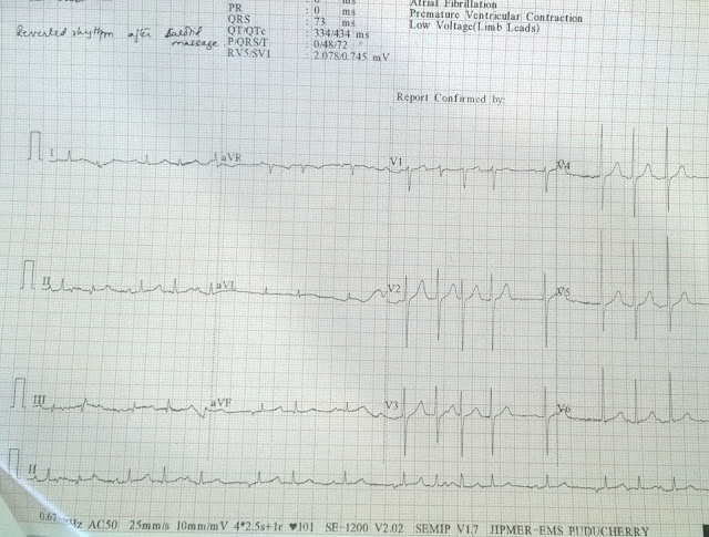
A 76 years old male resident of Thiruvannamalai, Tamilnadu,  came to JIPMER hospital Pondicherry with chief complaints of acute onset palpitation since last one hour, which was acute in onset, no associated with any chest pain, giddiness or vomiting. There was no history of diabetes mellitus, hypertension, coronary artery disease or smoking. During evaluation in the emergency department patient pulse rate was 190 beats per minute, BP-110/60 mmhg. ECG of the patient done in the emergency department, which is shown below.

                                                                      ECG 1 (Click on the image to enlarge it)


Description of the ecg-Narrow QRS complex regular tachycardia with heart rate aound 190 beats per minute, P wave are negative in lead II, III,avF and there is Pseudo R wave seen in lead V1, QRS alternans is present, no other ST-T wave changes were present. RP interval 140 msec, PR interval 200 msec so the ecg is suggestive of Short RP tachycardia so the diagnosis is PSVT with differential diagnosis being AVRT first and AVNRT second.

Patient was given carotid sinus message and his rhythm converted to sinus rhythm.


Sinus ECG of the patient-Sinus rhythm at rate 120 beats per minute, normal axis, no ST-T wave changes seen.

Patient underwent electrophysiological study at JIPMER hospital which was suggestive of AVRT with concealed accessory pathway.

                             Lets see how to approach in a patient with Narrow QRS Complex tachycardia


Thank you.


Severe sinus node dysfunction in Old age male

A 83 year old male resident of Pondicherrry, South India came to the JIPMER hospital emergency department with  chief complaints of one episode of giddiness along with vomiting today afternoon. There is history of dyspnoea along with palpitation were present but there was no history of chest pain, perspiration, or loss of consciousness. Patient was a known case of diabetes mellitus, hypertension since last 20 years and was on regular medication. Patient was also a known case of coronary artery disease with triple vessel diseases for which he underwent coronary artery bypass graft in 2011 at private hospital. During evaluation in the emergency department patient pulse rate was 24 beats per minute, BP-140/80 mmhg. ECG done in the emergency department is shown below.

                                                                       ECG 1


Description of the ECG-Junctional escape beats at rate around 18 beats per minute present, escape beat is of RBBB morphology with QRS duration 120 msec,single sinus beat is visible with visible P wave, PR interval 200 msec, with QRS of RBBB morphology, QT interval nearly 520 msec QTc 285 msec, so the ecg is suggestive of severe sinus node dysfunction with sinus arrest. 

                                                                                ECG


Another ECG of the patient again showing severe bradycardia with sinus pause with feature suggestive of  severe sinus node dysfunction.

ECG of the patient was again repeated after ten minute and cardiac marker of the patient were sent.

                                                                                     ECG 3


Description of the ECG-Normal sinus rhythm at 80 beats per minute, normal axis, PR interval 160 msec, complete RBBB, ST elevation was present in lead V1-V4 but there were no reciprocal change in inferior lead (II,III,avF), and as compared to old ecg there was no fresh ST-T segment changes. 

Patient cardiac marker, Trop I was within normal limit. So in view of intermittent sinus node dysfunction patient was decided to put on holter monitor to confirm the diagnosis. 
Holter monitoring of the patient done, which is shown below


Holter monitoring of the patient shown sinus node arrest with sinus pause more than 3 seconds. Maximum pause was of 4.032 sec, so in view of severe sinus node dysfunction permanent pacemaker was inserted and patient was discharged in stable condition.

Thank you.


Tuesday 17 January 2017

Middle age male with tachycardia. What is your diagnosis?

A 43 year old male came to JIPMER hospital, Pondicherry, India with chief complaints of palpitation on and off since last 2 years which were acute in onset, not associated with  any exertion, vomiting, nausea, vomiting, giddiness, chest pain or syncope. Tachycardia subsided on its own after one or two hours. There was no history of diabetes mellitus, hypertension, hyperthyroidism, coronary artery diseases. Last episode of palpitation occur 25 days back, ECG done at private hospital following which patient was referred to JIPMER Cardiology department for electrophysiological study and further managment. During evaluation in the OPD patient pulse was 94 beats per minute, BP 146/90 mmhg, cardiovascular and respiratory system were unremarkable. ECG of the patient is shown below.


ECG during tachycardia


Continued.................


Description of the ECG-Narrow QRS complex tachycardia with heart rate 250 beats per minute, normal axis, Negative P wave seen in lead II,III,avF, Pseudo R wave seen in lead V1, RP interval nearly 50 msec, PR interval 200 msec, no ST-T wave changes, no QRS alternans seen so the ecg is suggestive of Narrow complex regular short RP tachycardia most likely etiology being AVNRT.


ECG during sinus rhythm


Normal sinus rhythm at 75 beats per minute, normal axis, no ST-T wave changes seen.

So the diagnosis is PSVT most likely AVNRT. Lets see how to approach in a patient with narrow complex regular short RP tachycardia.


Thank you.

Monday 16 January 2017

ECG-Broad complex tachycardia in middle age male ??Diagnosis

A 45 years old male came to JIPMER hospital pondicherry, India with chief complaints of palpitation since last four days, which was acute in onset associated with one episode of vomiting, intermittent to continuous in nature, associated with atypical chest pain and breathlessness. Patient was a known case of diabetes mellitus. Patient also giving history of Old acute anterior wall myocardial infarction in february 2015, for which coronary angiography done at  JIPMER hospital was suggestive of recanalized Left anterior descending coronary artery. Patient lost to followup and stopped all medication since last six months. During evaluation in the emergency department patient pulse was 200 beats per minute, BP-100/60 mmhg, CVS S1S2 were present, Respiratory system bilateral equal air entry and there were no crepitation. ECG of the patient done in the emergency department is shown below.
   

ECG of the patient during tachycardia


 ECG Continued-----


ECG-Broad complex regular tachycardia with rate around 215 beats per minute, axis being left axis deviation, QRS duration in lead with maximum wide QRS in lead V1 is 240 msec, QRS being LBBB morphology, P wave are seen merged in QRS complex suggestive of AV dissociation, but there were no capture beat, fusion beat so the diagnosis of the ecg is most likely ventricular tachycardia with Supraventricular tachycardia with aberrancy being second differential diagnosis.Patient was started on injection amiodarone. Patient tachycardia reverted with injection amiodarone. ECG after injection  amiodarone is shown below.


ECG after giving injection amiodarone


Baseline ECG after giving injection amiodarone is suggestive of normal sinus rhythm with rate around 83 beats per minute,left axis deviation,narrow QRS complex, no preexcitation seen, intermittent ventricular premature contraction seen

So the final diagnosis is Ventricular tachycardia. Lets us see how to approach in a patient with broad complex tachycardia


Thank you.


Broad complex tachycardia in female ?Diagnostic dilemma

A 37 year old female came to JIPMER cardiology OPD with chief complaints of two episode of  palpitation over last one year. Patient went to private hospital and DC cardioversion was done. Patient was a known case of diabetes mellitus since last one year and was on dietary management. Patient was also giving  history of inferior wall myocardial infarction in 2015, for which she was treated at private hospital with thrombolytic therapy with tenecteplase and was started on medical management.Patient was doing well till  she developed one episode of tachycardia in october 2016 for which she was cardioverted with DC shock and was started on medical management. But again on  november 2016(14/11/2016) she developed one episode of tachycardia for which she was DC cardioverted. Later patient underwent coronary angiography (15/11/2016) which was suggestive of spontaneous dissection of right coronary artery from proximal to mid part with right dominance, rest of the coronary artery were normal. Following this patient was  referred to JIPMER Hospital, Pondicherry for electrophysiology study and further management.During evaluation at JIPMER hospital patient Pulse was 54 beats per minute, blood pressure 110/70 mmhg, ECHO done which was suggestive of hypokinesia of inferior wall, inferoseptal, posterior wall with  moderate mitral regurgitation, LVEF was around 50%. 

Previous ECG of the patient during tachycardia is shown below(Click on the image to enlarge it)


ECG-Broad complex regular tachycardia with heart rate around 200 beats per minute, QRS duration around 200 msec, QRS morphology being RBBB,  right axis deviation,no obvious P wave seen. There were no capture beat,fusion beat, QR wave seen in lead V1, RS wave seen in lead V6 with R/S ration being less than one, large R wave seen in lead avR, so this is a broad complex regular tachycardia with differential diagnosis being First Ventricular tachycardia second being Supraventricular tachycardia with aberrancy.


Another ECG of the same patient during tachycardia


ECG is suggestive of broad complex regular tachycardia with RBBB morphology with right axis deviation.


ECG of patient during sinus rhythm


ECG Normal sinus rhythm at 50 beats per minute, normal axis, QS complex in lead II,III,avF, no other ST-T wave changes seen.

So in view of baseline ecg suggestive of narrow QRS complex with past history of myocardial infarction patient ecg diagnosis being ventricular tachycardia. Patient underwent electrophysiology study followed by CARTO guided ventricular tachycardia ablation at JIPMER cardiology department by Dr Raja Selvaraj successfully. Presently patient is planned for OCT guided PCI to RCA in view of her coronary angiography suggestive of RCA dissection.
Now lets discuss how to approach in a patient with broad complex tachycardia. Kindly see the flow diagram given below. (Click on the image to enlarge it)


Thank you.


ECG-Tachycardia in young female ??What is your diagnosis

A 38 year old female resident of Pondicherry, India came with chief complaints of palpitation on and off since last one year. Last episode of palpitation occur 15 days back, which were acute in onset, associated with atypical chest pain but there was no  associated nausea, vomiting, giddiness or syncope, it was of one hour duration following which patient was admitted in private hospital, diagnosed to have abnormal rhythm during ecg evaluation. Tachycardia of patient was rellived by intravenous adenosine 6 mg. Patient was referred to JIPMER hospital, Pondicherry, India for electrophysiology study and further management.


ECG 1(Click on the image to enlarge it)


Description of the ECG-Narrow QRS complex, regular tachycardia with heart rate around 210 beats per minute, Negative P wave seen in lead II, III, avF, with Pseudo R wave seen in lead V1, RP interval 60 msec, PR interval 280 msec, no significant ST-T wave changes seen, normal axis. So the ecg is suggestive of Short RP narrow complex regular tachycardia suggestive of paroxysmal supraventricular tachycardia.


ECG after giving injection Adenosine 

Normal sinus rhythm at 100 beats per minute, normal axis, no pre-excitation seen, no ST-T wave changes seen.

So on the basis of these two ecg the diagnosis of this patient is Paroxysmal supraventricular tachycardia (PSVT),most likely AVNRT. Let discuss how to approach in a patient with narrow complex regular tachycardia.


Thank you.


First degree AV block associated with acute anterior wall myocardial infarction

A 48 years old male comes to JIPMER hospital, Pondicherry with chief complaints of acute onset retrosternal chest pain of three hours duration along with breathlessness. There was no history of nausea, vomiting, giddiness. Patient was a known case of chronic smoker since last 40 years and used to smoke 10 cigarettes daily. There was no history of diabetes mellitus, hypertension, past history of coronary artery disease, or family history, or dyslipidemia. During evaluation in the emergency department patient pulse was 50 beats per minute, cardiovascular system was normal, Respiratory system was normal and there were no crepitation or rhonchi. ECG of the patient done in the emergency is shown below.

ECG 1 (Click on the image to enlarge it)
 Description of the ECG-Normal sinus rhythm at 54 beats per minute, normal axis, PR interval was significantly prolonged with PR interval being 280 msec, narrow QRS with ST segment elevation in lead V2-V5, I, avL, with ST segment depression in lead III,avF so the ecg was suggestsive of acute anterior wall myocardial infarction with first degree AV block.

Patient was immediately thrombolysed with streptokinase, post thrombolysis ecg at 90 minute  is shown below.


                                                           ECG 2(Kindly compare this ecg with ECG 1)

Description of the ECG-Normal sinus rhythm with rate 75 beats per minute, PR interval now 200 msec, there is resolution of ST segement elevation by more than 50% in all the lead (V2-V5, I,avL), although resolution of ST segment in lead V4 is less as compared to other lead. QS complex were present in lead V1-V3, few Ventricular premature complexes are also present. So patient underwent successful thrombolysis.