Saturday, 13 May 2017

Friday, 12 May 2017
PSVT
A 21-year old female came with history of palpitation on and off since last five year.
ECG 1 (Click on the image to enlarge it)

ECG is showing narrow qrs complex regular tachycardia. I can not see any clear P wave or probably few doubtful P wave merged in the qrs complex seen at the end of qrs in lead II. So it is a short RP tachycardia. There were no ST- T wave changes.
ECG after giving adenosine injection (Click on the figure to enlarge it)

ECG is showing normal sinus rhythm at 75 beats per minutes, PR interval 130 msec, no evidence of any delta wave or any pre-excitation any where seen in the ecg. There are no ST-T wave changes.
So the final diagnosis of this patient is Short RP tachycardia and differential could be AVNRT, Atrial tachycardia or AVRT. Because P wave are merged into the qrs complex or very near the QRS complex(if i take negative complex seen in figure 1 in lead II), then the RP interval is less than 70 msec so most likely diagnosis is AVNRT.
Patient underwent Electrophysiological study and it show dual AV node physiology with AVNRT. Patient underwent successful radiofrequency ablation of slow pathway. Post ablation there is no evidence of any tachycardia. Patient was discharged in stable condition.
Let's discuss how to approach in a case of Narrow QRS complex tachycardia.

Thank you.
Thursday, 4 May 2017
Anterior wall myocardial infarction
A 52-year-male came to JIPMER hospital with history of acute onset retrosternal chest pain since 4 hours of duration along with palpitation, perspiration, giddiness, dyspnoea. There was no history of orthopnea, PND, syncope. His BP was 140/80 mmhg, Pulse 104 beats per minute, saturation of oxygen was 96%. Patient serial ECG were done which are shown below.
ECG 1

ECG is showing normal sinus rhythm at 100 beats per minute, normal axis, normal PR interval (120 msec), there is mild ST segment depression in lead I,avL (1 mv), there were no other significant ST-T wave changes seen.
Patient was kept under constant observation. His cardiac marker were sent and he was started on antiplatelet therapy along with heparin. But patient chest pain did not improve and suddenly he develop ST elevation acute anterior wall myocardial infarction after 3 hours of presentation.
ECG 2 (3 after presentation)

ECG is showing ST elevation in lead V2-V6 with hyperacute Tall T wave. Also there is mild ST segment elevation in lead I, avL.
ECG 3

ECG is showing ST elevation extensive anterolateral wall myocardial infarction. Patient was immediately started on streptokinase therapy. His post thrombolysis ECG is shown below
ECG 4(Post thrombolysis)

ECG 5 (Post thrombolysis)

ECG is showing qRBB with QS complex in lead V1-V5, avL suggestive of failed thrombolysis in this patient.
So the final diagnosis is Acute extensive anterior wall myocardial infarction with failed thrombolysis.
Thank you.
Wednesday, 3 May 2017
Inferior wall myocardial infarction presenting as complete heart block to Type II AV block
A 50 year old male presenting with history of giddiness along with 3 episode of vomiting since 4 hours of duration. There was no history of chest pain, palpitation, sweating, syncope. Patient was non diabetic, non hypertensive. His pulse was 50 beats per minute, BP-110/70 mmhg during the time of admission. Patient ecg done in the emergency department is shown below.

ECG of the patient is showing complete AV dissociation with ST segment elevation in lead III, avF, but elevation was less than 1 mv......


ECG is showing ST segment elevation in lead III, very mild ST segment elevation in lead II, avF

ECG is showing ST segment elevation in lead II,III, avf, with hyperacute Tall T wave, now instead of complete AV block there is 4:3 type I 2nd degree AV block (Wenckebach block) with group beating present.

ECG is not showing any evidence of right sided infarction as there is absence of ST segment elevation in right sided lead.
Patient was immediately started on thrombolysis.

ECG is showing complete resolution of ST segment along with resolution of AV block. PR interval is although borderline prolonged, PR interval after thombolysis is 160 msec.

ECG become completely normal next day. There is no evidence of AV block, neither any evidence of ST segment elevation.
Next day patient underwent coronary angiography which shows 70-80% stenosis in Mid right coronary artery(RCA) rest of the vessel were normal.
So the final diagnosis of the patient is IWMI with complete heart block with successful thrombolysis with Single vessel diseases of RCA.
Thank you.
ECG 1 (Click on the image to enlarge it)

ECG of the patient is showing complete AV dissociation with ST segment elevation in lead III, avF, but elevation was less than 1 mv......
ECG 2 taken after 10 min

ECG is still showing same finding as in first ECG
ECG 3

ECG is showing ST segment elevation in lead III, very mild ST segment elevation in lead II, avF
ECG 4 done after 30 min

ECG is showing ST segment elevation in lead II,III, avf, with hyperacute Tall T wave, now instead of complete AV block there is 4:3 type I 2nd degree AV block (Wenckebach block) with group beating present.
ECG with right sided lead
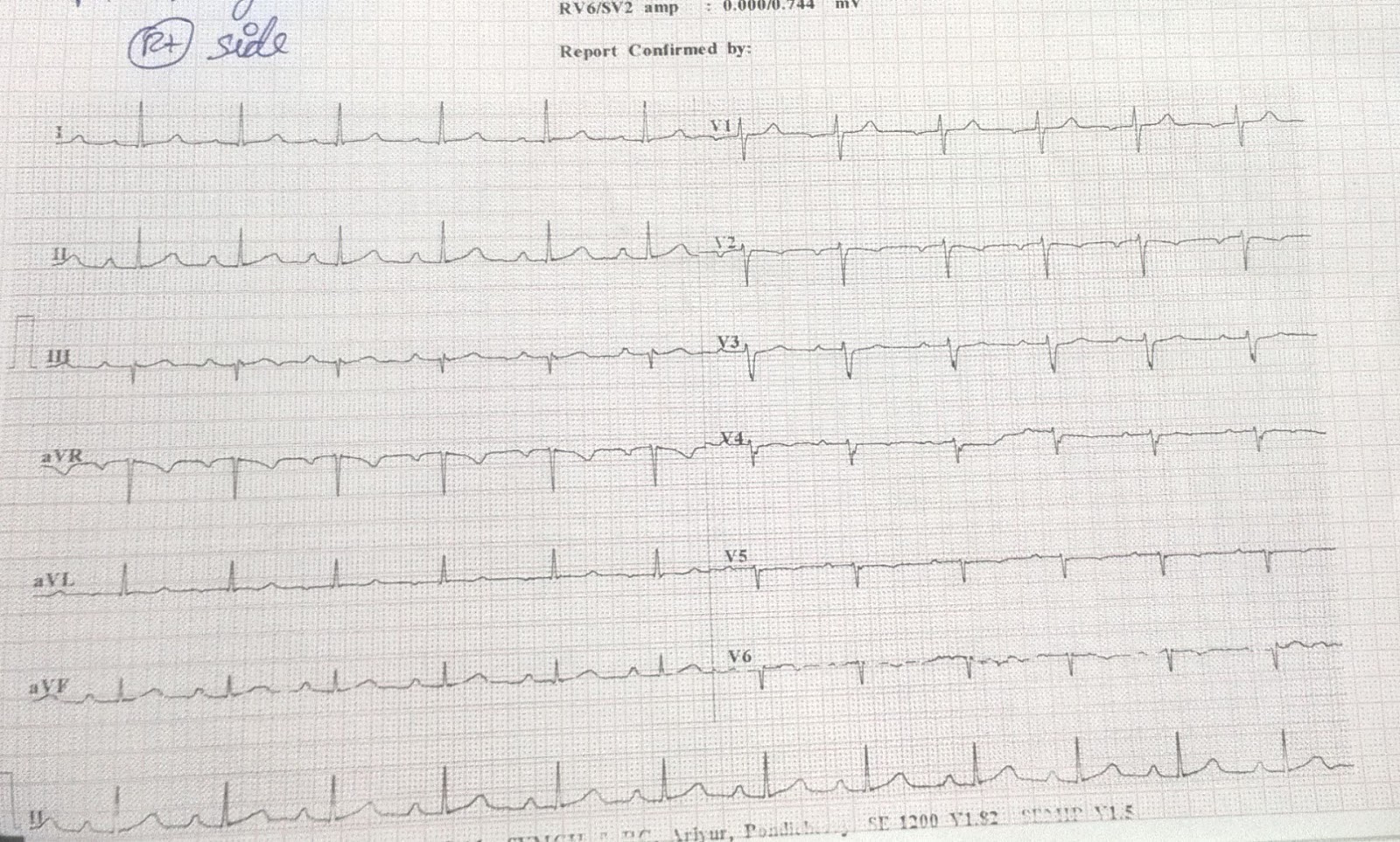
ECG is not showing any evidence of right sided infarction as there is absence of ST segment elevation in right sided lead.
Patient was immediately started on thrombolysis.
Post thrombolysis ECG is shown below
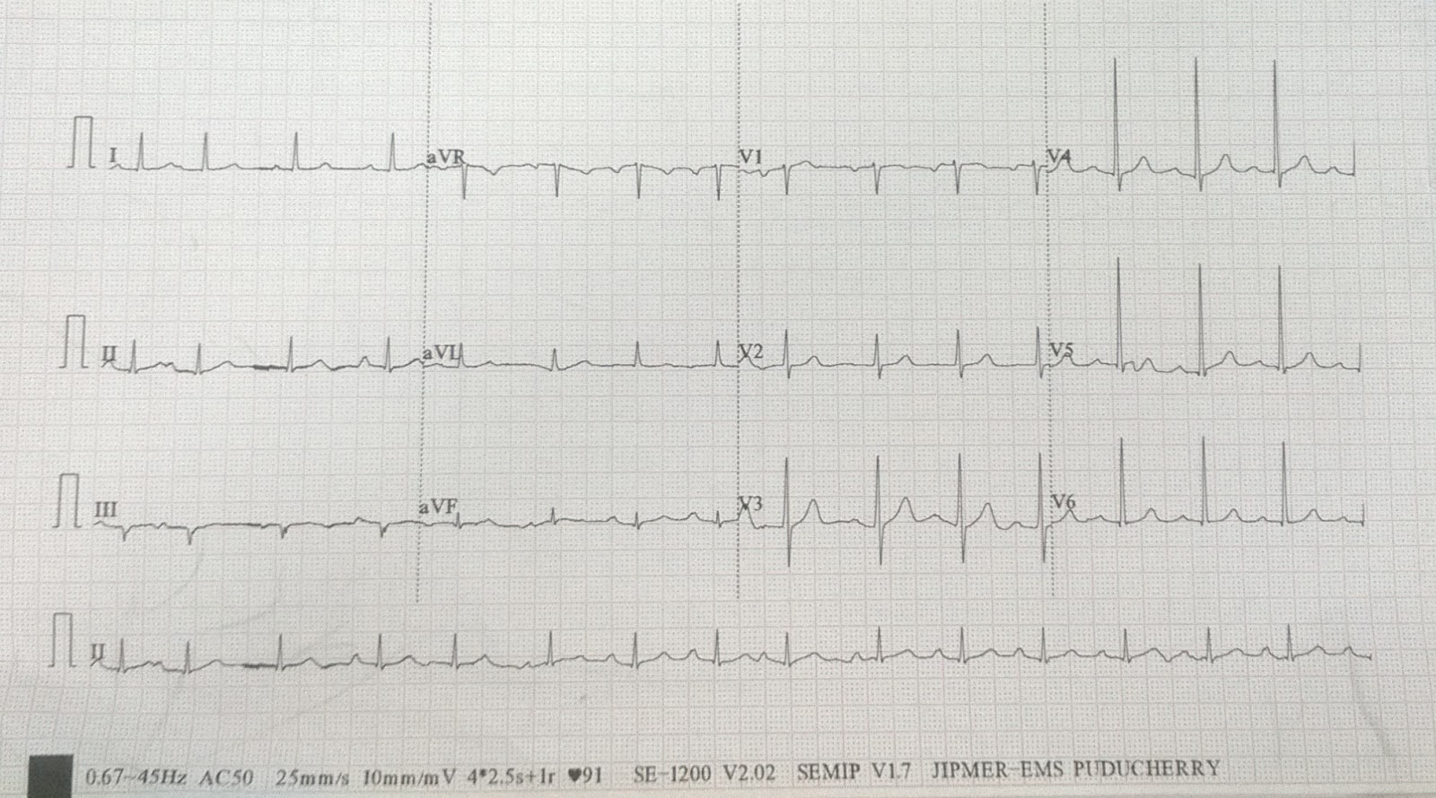
ECG is showing complete resolution of ST segment along with resolution of AV block. PR interval is although borderline prolonged, PR interval after thombolysis is 160 msec.
ECG done next day

ECG become completely normal next day. There is no evidence of AV block, neither any evidence of ST segment elevation.
Next day patient underwent coronary angiography which shows 70-80% stenosis in Mid right coronary artery(RCA) rest of the vessel were normal.
So the final diagnosis of the patient is IWMI with complete heart block with successful thrombolysis with Single vessel diseases of RCA.
Thank you.
Middle age female came with palpitation ??? what is your diagnosis
A 50-year-old female came to JIPMER hospital with chief complaints of acute onset palpitation since last 1 hours , which were acute in onset was not associated with nausea, vomiting, giddiness or syncope. Patient was giving similar history since last on and off since last 2 years. Patient was non diabetic, non hypertensive. Her tachycardia ECG is shown below.
ECG 1(Click on the image to enlarge it)

ECG of the patient is showing narrow complex regular tachycardia at heart rate of 188 beats per minute, normal axis, QRS duration 80 msec, P wave have merged into the T wave and producing ST segment depression in lead I,avL, II,III,aVF, V5,V6. Kindly see lead III, avL, i think those are the inverted P wave so the RP interval is 160 msec and PR interval is 200 msec, so it is short RP tachycardia.
ECG 2(Showing Precordial lead)

Precordial lead of the patient is showing narrow complex, regular, short RP tachycardia.
So the differential diagnosis in this patient is either AVRT or AVNRT. Because the RP interval is more than 70 msec so the possibility of AVRT was more compared to AVNRT.
In view of her continuous palpitation initially vagal maneuver was tried but it was not successful so patient was given intravenous adenosine 6 mg via antecubital vein. Her tachycardia subsided after giving adenosine. Her sinus ecg is shown below.
ECG after giving injection adenosine

ECG is showing normal sinus rhythm at 73 beats per minute, normal PR interval, no ST-T wave changes, normal axis.
Patient underwent Electrophysiological study which shows orthodromic AVRT with left accessory pathway. Interatrial septal puncture was done and successful ablation was done.
So the final diagnosis of this patient is Orthodromic AVRT with left sided pathway.
Now lets discuss how to approach in a patient with Narrow complex tachycardia.
Thank you.
Monday, 1 May 2017
Variable AV block, 2:1 AV block converting into 3:1 AV block
A 68-year old female came to JIPMER Hospital, Pondicherry, India with chief complaints of giddiness on and off since last 3 days along with dyspnoea on exertion NYHA II. Patient was a known case of hypertension and she was on beta blocker. ECG of the patient is shown below.
ECG 1(Click on the image to enlarge it)

ECG of the patient is showing bradycardia with heart rate around 50 beats per minute, normal axis, normal QRS duration, there are two P wave for every one QRS complex, PR interval is fixed, around 120 msec, so the ecg is suggestive of 2:1 AV block, it could be 2 nd degree Type 1 or Type II AV block. As the patient was on betablocker so the drug was stopped. Her ecg done after 2 days is shown below.
ECG after 2 days of stopping beta blocker

ECG is now showing 3:1 AV block, so the block had progressed instead of improving in spite of stopping beta blocker so it means this patient is having significant AV node diseases. In view of her ecg changes she was advised to undergone permanent pacemaker. Following this patient successfully underwent permanent pacemaker insertion (VVI).
Thank you.
WPW Syndrome
A 13-years-male came to JIPMER Pondicherry hospital cardiology OPD with chief complaints of palpitation on and off since last one year, history of syncope 3 episode over last 15 days along with atypical chest pain. ECHO of the patient was normal. ECG of the patient is shown below.
ECG 1(Click on the image to enlarge it)

Sinus rhythm at 90 beats per minute, left axis, PR interval 80 msec, Positive delta wave seen in leads 2,3,avf, negative delta wave seen in lead V1, ST segment depression present in lead I,II, III, avF, V5,V6. So the ECG of the patient was suggestive of WPW syndrome with Right free wall pathway.
In view of history of palpitation and syncope patient underwent Electrophysiological study which show Right free wall accessory pathway with preexcitation. Patient underwent successful ablation.
Post ablation ECG is shown below.
ECG 2 (Click on the image to enlarge it)

ECG is showing Normal sinus rhythm at 75 beats per minute, normal axis, no pre-excitation seen. There is Deep T wave inversion in lead V1,V2 also there is T wave inversion seen in lead V3 suggestive of memory T wave post ablation therapy.
WPW Syndrome
Algorithm for pathway localisation

Thank you.
Subscribe to:
Posts (Atom)