A 50-year-old female came to JIPMER hospital with chief complaints of acute onset palpitation since last 1 hours , which were acute in onset was not associated with nausea, vomiting, giddiness or syncope. Patient was giving similar history since last on and off since last 2 years. Patient was non diabetic, non hypertensive. Her tachycardia ECG is shown below.
ECG 1(Click on the image to enlarge it)
ECG of the patient is showing narrow complex regular tachycardia at heart rate of 188 beats per minute, normal axis, QRS duration 80 msec, P wave have merged into the T wave and producing ST segment depression in lead I,avL, II,III,aVF, V5,V6. Kindly see lead III, avL, i think those are the inverted P wave so the RP interval is 160 msec and PR interval is 200 msec, so it is short RP tachycardia.
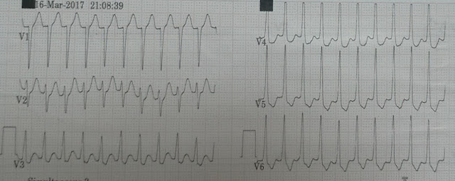
ECG 2(Showing Precordial lead)
Precordial lead of the patient is showing narrow complex, regular, short RP tachycardia.
So the differential diagnosis in this patient is either AVRT or AVNRT. Because the RP interval is more than 70 msec so the possibility of AVRT was more compared to AVNRT.
In view of her continuous palpitation initially vagal maneuver was tried but it was not successful so patient was given intravenous adenosine 6 mg via antecubital vein. Her tachycardia subsided after giving adenosine. Her sinus ecg is shown below.
ECG after giving injection adenosine
ECG is showing normal sinus rhythm at 73 beats per minute, normal PR interval, no ST-T wave changes, normal axis.
Patient underwent Electrophysiological study which shows orthodromic AVRT with left accessory pathway. Interatrial septal puncture was done and successful ablation was done.
So the final diagnosis of this patient is Orthodromic AVRT with left sided pathway.
Now lets discuss how to approach in a patient with Narrow complex tachycardia.
Thank you.


No comments:
Post a Comment